Case Study:
Displaced Flap Tears of the Medial Meniscus
Presented by: Dr Michael Goldberg
Presentation:
- A 46 year old man twisted his knee 2 months ago and felt immediate medial pain and a popping sensation. The knee swelled immediately.
- It was sore for a few weeks and then was starting to slowly improve.
- He had another twising episode one month later. He has had persistent medial pain since then, including in bed at night.
- Moderate knee joint effusion.
- Point tenderness medial joint line.
- Positive McMurray’s test.
- Cruciate and collateral ligaments are stable.
- Weight bearing xrays are normal.
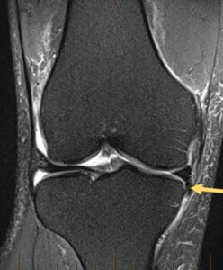
- MRI showed “a flap tear of the medial meniscal body with inferiorly displaced flap fragment into the medial recess.”
- Is this tear likely to improve with non-surgical management?
- Does this need specialist referral?
Answer:
It is sometimes difficult (even for Orthopaedic surgeons) to know which meniscal tears will respond well to conservative treatment, and which would do better with surgery. Patterns of meniscal tear which are best treated with surgery include bucket-handle tears, full thickness radial tears and meniscal root tears. Other tears, such as “complex” or “degenerative” tears are often less clear cut, and may improve with conservative management (anti-inflammatories, physiotherapy, activity modification).
Displaced meniscal flap tears occur when a fragment of torn meniscus displaces into the recess between the proximal tibia and the adjacent knee capsule and soft tissues. It commonly occurs after a defined traumatic incident (such as a twisting injury), but may also occur with no clear traumatic mechanism. Patients usually describe pain at the precise site of the tear and have point tenderness on examination. They often describe pain in bed at night, particularly when lying on their side with their contralateral knee putting pressure on the medial side of the affected knee. They often need to sleep with a pillow between their legs to alleviate this. Patients may or may not experience mechanical symptoms of locking and giving way.
This tear is usually best seen on the coronal T2-weighted MRI scan (see figure 1), where a fragment of meniscus (black in appearance) is stuck between the medial tibial plateau and the overlying medial collateral ligament.
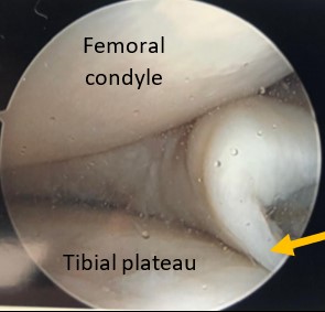
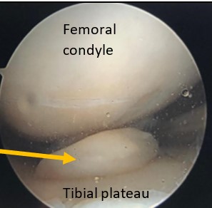
This tear pattern tends to be persistently painful, as the meniscal fragment becomes entrapped between bone and the adjacent soft tissues. Although a trial of conservative management is not unreasonable, in my experience, symptoms tend to persist and necessitate surgery. Surgery involves a knee arthroscopy, reduction of the displaced meniscal fragment from the medial recess (figure 2a & 2b) and debridement of the torn fragment. Patients who have arthroscopic debridement of these tears tend to be very happy after surgery, and usually get almost immediate and complete relief of their acute pain and tenderness.

Yellow arrow pointing to Medial meniscal fragment displaced into the medial recess. It is entrapped between the medial tibial plateau and MCL.

Using a probe to reduce the displaced meniscal fragment out from the medial gutter

Take home message: Consider early Orthopaedic referral for patients with displaced medial meniscal flap tears, as this pattern of tear tends to be persistently symptomatic, and patients usually
respond very well to arthroscopic debridement.


