Welcome to Question for Physiotherapists, October 2021.
This month Dr Todd Gothelf discusses Syndesmosis fixation.
Please feel free to send your questions to education@orthosports.com.au

QUESTION I I would like to understand more about syndesmosis fixation please. I remember seeing screws for fixation and then patients had to have them removed. I recently had a patient with a “tightrope” fixation with no plans for any removal. Can you please explain the differences in fixation for the syndesmosis and how they are treated differently?
ANSWER |
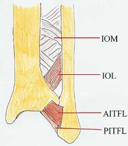
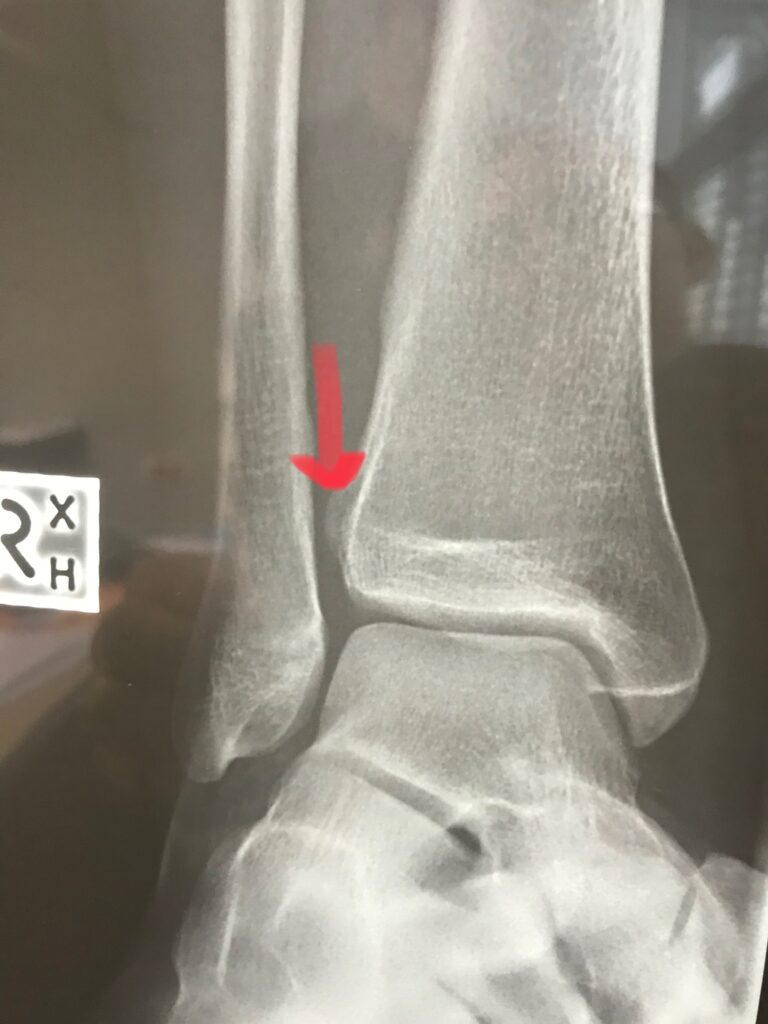
Background: The syndesmosis consists of the ligamentous structures that connect the distal tibia to the fibula and keep the fibula in the incisura fibularis. These structures include the Anterior Inferior Tib-Fib ligament (AITFL), the Posterior Inferior Tib-Fib ligament (PITFL), Interosseous Ligament (IOL) and the interosseous membrane (IOM). A rupture of these structures, even without fracture of the fibula, can result in instability between the tibia and fibula and allow lateral movement of the talus. Studies have shown that with as little as 1mm of lateral displacement of the fibular, the tibiotalar contact area available for weight bearing is reduced by 42%. A lack of anatomic reduction of the syndesmosis can result in increased stresses across the joint, resulting in early degeneration or arthritis.
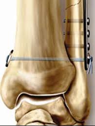
Fixation: With an acute syndesmosis rupture, surgical strategy involves reducing the fibula to its proper relationship to the tibia, and fixing the position. The traditional fixation for a syndesmosis was screw fixation. A screw would fix the position of the fibula to the tibia to allow the ligaments to heal. After adequate healing of the ligaments, around three months, the screw is removed to allow the natural micromotion of the ankle joint again. Screw fixation is still commonly used. Screw fixation is reliable in holding the reduction of the syndesmosis. A theoretical disadvantage is that the construct is too stiff and non-physiologic. A further disadvantage is that a second surgery is usually needed to allow more natural movements. During recovery the screw or screws may break.
More recently a suture button fixation has been used and has gained popularity. The fixation is flexible with multiple strands of non-breakable suture across the joint. The theoretical advantages are that suture fixation allows for more physiologic motion between the tibia and fibula. Because the construct is flexible, most surgeons leave the fixation in place avoiding the need of a second surgery. Studies have shown that the suture fixation is similar in strength to screw fixation.
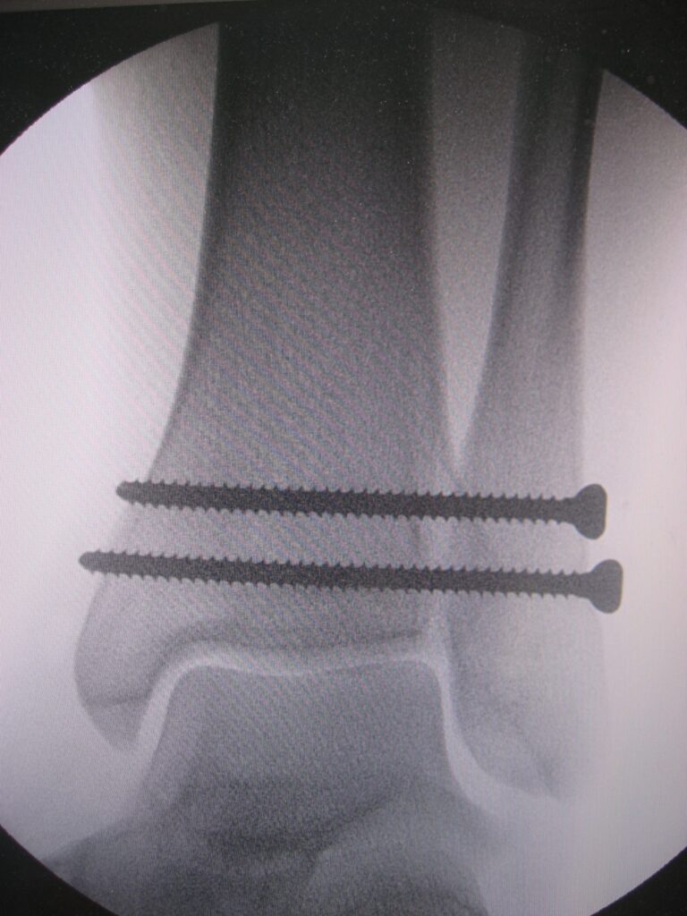
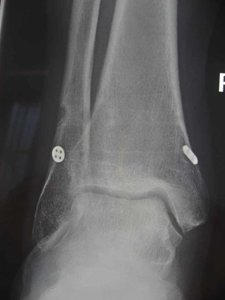
With either screw fixation, or suture fixation, the post-operative instructions are usually similar. Treatment usually involves non-weightbearing with Camboot immobilisation for six weeks, followed by weight bearing in a Camboot for four weeks. Ankle motion is commenced at six weeks after surgery and progression with strengthening and return to activities after three months from surgery. The main difference in treatment is that the tightrope is often left in place, while screws are usually removed by a second operation at three months from surgery.

