This month Dr Doron Sher presents a Knee Examination series that you may find useful for patients presenting in your practice requiring a knee assessment. Please email us with any questions you may have to : education@orthosports.com.au
Knee Examination Series:
Presented by: Dr Doron Sher
To view the Knee Examination Video please Click Here
A careful history offers a high index of suspicion to the diagnosis for most knee injuries. This includes previous injuries, the mechanism of injury, the development of pain, swelling, instability, locking and the response to treatment.
Meniscal Tears:
History – meniscal tears may present acutely or as overuse. Localised joint line pain, mechanical symptoms (locking or catching) and movement restrictions are all features.
Gait and stance – the patient may have a limp , limited movement or pain squatting.
Effusion – You do not have to have a swollen knee to have a meniscal tear. There will always be swelling with a chondral injury but not always with a meniscal tear.
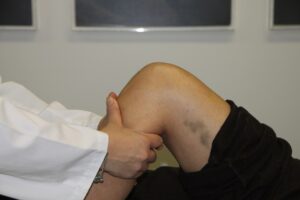
Joint line tenderness – useful but not very specific. The knee is bent to 90 degrees and the entire joint line is palpated looking for pain. Allowing the hip to drop out the side will allows more specific palpation the medial meniscus.
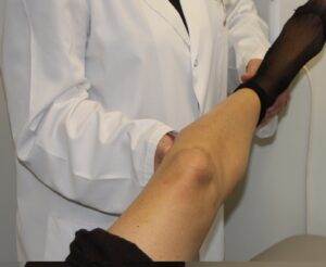
McMurray’s Test – With the patient supine, bend the knee and twist the foot (internal and external rotation). A click (and often pain) is felt at the joint line as the knee is brought into flexion.
ACL tears:
Anterior cruciate ligament tears present as acute injuries. This may be contact or non-contact. In the case of non-contact injury the history is usually of a patient playing sport with the foot planted attempting a side stepping manoeuvre. The knee gives way with a popping sound and the patient falls to the ground. Occasionally the patient may describe a hyperextension injury or a quadriceps active mechanism when the pop occurs as they jump into the air. The knee may swell immediately and the gait painful. Most sportspeople are unable to continue playing their sport at the time of injury.
On examination it is important to ensure that the patient is relaxed. This is often difficult when the knee is painful and swollen and the examination can be challenging. In order to obtain relaxation, the patient is asked to rest his or her head on a pillow with their arms by their sides. It may be useful to gently roll the thigh in and out, to get the muscles to relax. Placing a pillow below the knee is often more comfortable for the patient.
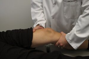
Lachman Test: The knee is unlocked in 30° of flexion. The patient’s heel rests on the couch. The examiner holds the patient’s tibia, with the thumb on the tibial tubercle. The examiner’s other hand is placed on the patient’s thigh, a few centimetres above the patella. Placing your leg under the thigh can make it easier to control the femur in larger patients. The hand on the tibia applies a brisk anteriorly directed force to the tibia.

The quality of the endpoint at the end of the movement is described as either “firm” or “soft “ and is always compared to the other knee. If the movement of the tibia on the femur comes to a sudden stop, this is described as a firm endpoint. A soft endpoint almost always indicates a torn ACL. If the ACL is torn in one knee, the patient will usually be aware of the difference between the firm endpoint in the healthy, and the soft endpoint in the cruciate-deficient knee. A firm endpoint results from the sudden tensioning of the ACL. Some patients are able to discriminate the side-to-side differences in the quality of the endpoint.
Active resisted extension
In obese patients, subjects with bulky muscles or patients with a large effusion, it may be difficult for the examiner to encircle the patient’s thigh with his or her hand. In such cases, the examiner may place a fist under the knee, hold the ankle against the couch with the other hand, and ask the patient to lift the leg against resistance. This resisted quad setting will move the tibial tubercle forward. This is a useful screening test but takes some practice to appreciate the anterior translation of the tibia which indicates the torn ACL.
Pivot shift: Tests screening for pivot shift were first described by M. Lemaire, in 1968. Since then, many such tests have been devised: A shift means that the ACL has gone. Sometimes, though, the ACL may be deficient without a pivot shift occurring.
The pivot shift test of MacIntosh “When I pivot, my knee shifts.” This is how a hockey player described his symptoms and so a test was devised to reproduce this sensation. It involves stress applied to the knee in valgus and flexion, with or without internal rotation.

Description of the test: The patient is positioned supine and you stand on the affected side. You use one hand to hold the patient’s foot in very slight internal rotation. With the other hand, you apply a valgus stress to the posterolateral aspect of the proximal calf. At this point, flexion is started. The lateral tibial plateau will be seen to sublux forwards during the first degrees of flexion. As flexion progresses, the anterolaterally subluxed tibia will suddenly reduce, at 30° of flexion. This reduction is associated with a characteristic clunk, which the patient will readily recognise. (If you wish to gain expertise with this test, you may find it helpful attending one of our knee surgery lists and examine the knees of patients under anaesthesia).
Anterior drawer in 90° flexion or direct anterior drawer
The examiner sits on the patient’s foot, which has been placed in neutral position. The knee is in 90° flexion. The index fingers are used to check that the hamstrings are relaxed, while the other fingers encircle the upper end of the tibia and pull the tibia forwards.

If a direct anterior drawer is obtained, the ACL will be torn. However, for this sign to be elicited, peripheral structures such as the medial meniscus or the menisco-tibial ligament must also be damaged. This ligament forms a wedge, in 90° flexion, preventing anterior tibial translation. The finding of an anterior drawer is conclusive evidence of an ACL tear. However, not every ACL tear will be associated with a positive anterior drawer test.


