Tip Toe Walking
Walking on tip toes is a common postural variation which causes parental concern. One can essentially divide these children into four groups:
Habitual Tip Toe Walkers
This is by far the largest group – usually children between 2 and 5 years of age who have always walked on tip toe. They can commonly stand with their feet flat and may be able to walk on heels but walk on tip toes out of habit. The angle can be corrected above a right angle. These children have a normal birth and development history.
Neuromuscular Diseases
Cerebral palsy (spasticity) will cause children (and adults) to walk on tip toes. This is because their calf muscles and Achilles tendon contract because of abnormal nerve supply and consequent abnormal growth. There may be a history of a difficult birth or pregnancy. Reflexes are increased and walking may be delayed. Treatment involves physical therapy and sometimes surgery.
Muscular dystrophy
Walking on tip toes is occasionally the presenting sign in young boys with Duchenne’s muscular dystrophy. There will usually be signs of muscle weakness and enlarged calves. A serum blood test for CPK (muscle enzyme) level is recommended.
Structural Tip toe Walkers
A very small number of children have a true contracture of their Achilles tendon without any other cause demonstrated. This is very rare and if marked, surgical lengthening of the tendon may be needed in the older child. In this group, the ankle can’t be corrected to a right angle or beyond.
The vast majority of children who present with tip toe walking will be in the habitual group and the posture should resolve with time. Casting, exercises, special shoes and orthotics have not been demonstrated to make any difference to this group.
In-Toeing And Out-Toeing In Children
In-toeing and out-toeing occur in children and are often considered variations of normal rather than abnormalities. In-toeing (commonly called pigeon toeing) is the tendency for the toes to point inwards. It is possible to have one foot turning in and the other turning out.
These conditions are usually the result of torsion (twisting) of the bones in either the foot, the lower leg or around the hip. Most of these are the result of positions the body adopts in the uterus. Sometimes it can be an inherited condition.
Many fine athletes have been “in-toers” as youngsters, so the condition does not generally cause difficulties with sports. It does not lead to arthritis in adulthood.
Some postural abnormalities may be the result of conditions that imply more serious problems. Deep skin creases in the foot, one foot being noticeably smaller than the other, and under-developed calf muscles may indicate a serious condition. Your family doctor can help decide if your child has a condition that will need orthopaedic advice or not.
Treatment
You now know that a child who turns in or out at the foot may have a twist either in the foot, the lower leg or the hip. Not infrequently, the torsional deformities can be at one or more level.
A child who has a foot that is twisted inwards is said to have metatarsus adductus. In this condition the bottom of the foot (sole) assumes the shape of a banana. It is important in this condition to differentiate the relatively harmless condition of metatarsus adductus from the more serious condition of congenital club foot. The majority of metatarsus adductus will correct even without treatment and generally by the time the child is 5 years of age. A small percentage (of up to 20%) may be left with a variable degree of twisting of the foot after the age of 5. Generally, if the deformity is severe, the condition is best treated early. Passive stretching may be recommended, as may the use of “straight-last” shoes which hold the foot in a corrected position.
Children who turn in at the lower leg level are said to have internal tibial torsion. This condition is quite common in children under 18 months of age. It usually resolves itself with further growth and only very occasionally does it require treatment.
In a child over 18 months of age, a twist in the thigh bone at the hip level is the most common of in-toeing. This condition is commonly called “inset hips”. It is known as persistent femoral anteversion. The condition usually resolves spontaneously, though it may take up to the age of 10 years for it to correct completely.
More than 90% of children with this condition often prefer to sit with their hips twisted inwards in a “W”. Whilst this is not the cause of the condition it can hinder spontaneous correction of the deformity. Children with this condition are, therefore, advised to sit cross-legged with their legs in front of them.
Very occasionally a child with inset hips is seen to have a significant degree of deformity persisting after the age of 10. Where the deformity is severe after this age, surgery may occasionally be needed to correct the deformity. This situation does not arise very often at all and in 99% of cases no surgery is necessary.
Children with pigeon toe often look clumsy when they run or when they are tired. This is not a matter for great concern as it will correct when the deformities resolve.
Children whose feet turn out, may do so also from a number of causes. Often times it is a turning out of the foot/ankle level. These children are often a little flat footed and this is often an inherited phenomenon. The young baby who has his leg turned out may do so at the feet level and often also from the thigh level. A turning out of the thigh bone is a very common finding in a young baby. This infantile position will correct itself, generally within a couple of years. It requires no active treatment.
Where a child has a leg that is short as well as turned out, the possibilities of a dislocated or under-developed hip joint should be considered. Such a child must be brought to see a doctor soon.
Shoes for Children
It is of little benefit to spend money on expensive shoes. Good shoes can be obtained at reasonable prices provided they fulfil the criteria below.
Shoes do not correct deformities. Poor fitting shoes, however, can cause problems with children’s feet as much as they do in adulthood.
A Checklist for Shoes
- Good fit – comfortably loose when worn with soft, absorbent socks.
- Shaped like the foot – broad and spacious in the toe area.
- Shock-absorbent sole – a low wedge type is best, never high heels.
- Breathable material – canvas or leather, not plastic.
Flatfoot
Flatfoot (pes planus or pes planovalgus) is common and rarely requires treatment.
Over 90% of infants have the appearance of flat-footedness. This is often due to a pad of fat which is normally situated on the inner surface of the foot and mostly resolves with growth. It requires no treatment.
About 10% of the population have true flat feet. There are basically two types, rigid and mobile.
Rigid flat feet are those which do not develop a normal arch when standing on the toes. There are many causes but all are uncommon. When rigid flatfoot is present it may cause pain and often requires treatment.
Mobile (or physiological) flatfoot is common and affects about 1 in 10 individuals. It is recognisable in that the foot arch reappears when the great toe is extended upwards and when standing on the toes. Mobile flatfoot is often familial and is generally regarded as a variation of normal. It often co-exists with knock knees (genu-valgum) and inset hips (persistent femoral anteversion).
Most children with flat feet are very supple and often have “ligamentous laxity”, which is a term meaning merely that they are more flexible than the rest of the population.
Mobile flatfoot is rarely painful and does not cause problems in later life. It is compatible with full sporting activities. Mobile flatfoot does not usually correct itself and is not influenced by the presence of arch supports and corrective shoes, despite what the manufactures say.
Shoes for Children
It is of little benefit to spend money on expensive shoes. Good shoes can be obtained at reasonable prices provided they fulfil the criteria below.
Shoes do not correct deformities. Poor fitting shoes, however, can cause problems with children’s feet as much as they do in adulthood.
A Checklist for Shoes
- Good fit – comfortably loose when worn with soft, absorbent socks.
- Shaped like the foot – broad and spacious in the toe area.
- Shock-absorbent sole – a low wedge type is best, never high heels.
- Breathable material – canvas or leather, not plastic.
Bow Legs
Genu varum is the normal physiological posture in the first two and a half years of life. It can persist into adulthood and may be associated with premature medial compartment osteoarthritis. There is some racial variation on the longitudinal alignment of the lower limbs with Asians tending towards genu varum.
The following features are suggestive of pathological bow legs…
- Deformity greater than 15 degrees.
- Asymmetrical genu varum.
- Lateral subluxation of the tibia.
- Recurvatum deformity of the knees or “back knee”.
- Other long bone deformities.
Possible pathological causes of genu varum include Blount’s disease, metabolic bone disease, skeletal dysplasia, post-trauma and infection.
Severe bow legs need to have the above causes excluded. Treatment if indicated usually consists of a genu varum brace which needs to be worn for at least 2 years. Surgery is occasionally indicated and usually consists of an upper tibial osteotomy, preferably after skeletal maturity.
Varus – bow legged.
Valgus – knock kneed (see below)
Knock Knees
Genu valgum is the normal physiological alignment of the limbs after the age of 3 years. Females tend to have greater valgus angulation in the knees than males.
Pathological causes of genu valgum include metabolic bone disease, skeletal dysplasia, infection, post-trauma and obesity.
Physiological genu valgum usually resolves to within the normal range by the age of 6 or 7 years. It can persist beyond this time and apart from being a cosmetically unattractive deformity it gives problems with patellar maltracking and patellar excess lateral pressure syndrome and recurrent dislocations.
Treatment is usually indicated if the inter-malleolar distance between the ankles with the knees together measures more than 10cm at the age of 10. Surgery is the best method of treatment and involves either stapling of the medial upper tibial and distal femoral growth plates before skeletal maturity or a supracondylar varus femoral osteotomy after skeletal maturity.
Heel Pain In Children
Pain in the heel is a common symptom in children between the age of 8 and 14.
It is commonly related to exercise and running. Epidemics occur at the start of soccer, football and athletic seasons.
The child complains of aching and sometimes limps and hobbles after sport. There may be local tenderness over the heel.
The problem is related to a stress/overuse injury which causes microdamage to the growth plate of the heel.
Treatment involves activity restriction, stretches for the Achilles tendon and the use of a shock absorbing heel insert.
X-rays are indicated in the presence of swelling, rest pain, night pain and severe local tenderness to exclude infection and tumour.
Perthe’s Disease
What is Perthes’ Disease?
This childhood condition is caused by disturbance of blood supply to the ball of the hip bone (femoral head). We do not know what triggers the condition.
The hip bone like all living tissue requires nutrition which it receives from blood that flows through fine blood vessels. In Perthes’ disease, a variable number of these vessels are blocked resulting in parts of the femoral head becoming non-viable (avascular necrosis).
Fortunately the child’s hip has great capacity to repair the damage. New blood supply will develop and repair of the damaged bone is usually complete in 2-3 years. This happens even if no treatment is given. The rate of repair is inversely proportionate to the age of the child, therefore younger children get over the condition quicker and have better outcomes.
The portion of the femoral head affected by avascular necrosis is mechanically weak and during the time it takes nature to heal the affected part, deformity may develop. The femoral head is normally spherical and fits perfectly with a cup (acetabulum) on the side of the pelvis to form the hip joint. This condition can cause the femoral head to flatten (non-spherical subluxation). The degree of decormity of the femoral head determines the outcome of the condition. Minor deformity is compatible with normal hip function but major deformity may result in premature arthritis of the hip joint.
Presentation
The condition affects children between the age of 4 and 10 years though older and younger children can be affected. It usually affects one hip but in 10% of cases both hips are affected.
The child with the condition develops a limp which worsens gradually. There is usually pain in the knee, thigh or groin when the child attempts to put weight on the leg or move the hip joint. The condition affects only the hip joint and there is no associated general illness.
Pain and limp is intermittent initially and becomes gradually more persistent. Mobility of the hip joint is reduced. If there is already some deformity of the femoral head, the affected leg is slightly shortened.
Diagnosis
The condition is diagnosed by x-rays in the established cases or by bone scans in the early cases. More recent investigations include MRI (magnetic resonant imaging) and ultrasonography.
Perthes’ disease can elude diagnosis in the early stages and may be confused with other conditions that also cause hip pain eg Transient synovitis, rheumatoid arthritis, septic arthritis.
Treatment
The aims of treatment are:
- Reduce hip irritability and stiffness.
- Prevent deformity of the femoral head.
- Protect the hip through the period of healing.
More than 50% of children affected by Perthes’ disease do very well without any active treatment. These are the children who acquire the disease when they are 5 years or younger and those in whom only a small portion of the femoral head is affected. These children require only regular checkups and occasional confinement to bed to treat hip pain and stiffness. Most doctors will advise against impact sports eg jumping and running on hard surfaces, for a period of two years.
Children who acquire the condition after 8 years of age may not benefit from active treatment. These children tend to develop more hip deformity and have less good outcome. They will nonetheless have hips that will function quite well for many years. There is a higher probability of hip arthritis after the age of 45 years.
Children with the condition between the ages of 5 and 8 years require careful evaluation to decide if they require treatment. Opinions amongst medical practitioners differ widely and there is, as yet, no generally agreed protocol for treatment.
Although all doctors agree that it is desirable to prevent hip deformity we cannot agree on how best to achieve this aim or even whether we can influence the development of hip deformity.
The majority of doctors involved in the care of children’s orthopaedics believe that femoral head deformity can be minimised or prevented by keeping the head under the protective cover of the acetabulum (Principles of Containment), provided treatment is carried out at an appropriately early stage of the disease. Containment can be by means of an abduction brace in which the head is kept under protective cover of the acetabulum by restricting motion within a selected range, or by surgically redirecting the femoral head (femoral osteotomy) or acetabulum (innominate osteotomy) to provide the cover. The difference in results of operative and non-operative treatment has not been conclusively determined. There are advantages and disadvantages with both methods of treatment.
The duration of treatment by bracing varies with the age of the child and to some extent the degree of head involvement. Braces are usually required for at least 2 years. If treatment by brace is selected, compliance to treatment is essential. If a child will not comply to brace treatment, operative treatment may be more appropriate.
An operation eliminates the need for constant supervision of the child, but one should be reminded that this option involves a significant and skilled surgical undertaking under general anaesthesia. The choice of operative technique is beyond the scope of this article but you are advised that the operation does not hasten healing of the condition.
A femoral osteotomy may cause slight shortening of the leg. An innominate osteotomy lengthens the leg slightly. It is unusual for either form of operation to require blood transfusion. Recovery from an operation involves variably 1 to 3 weeks of hospitalisation and use of crutches for about six weeks. Some surgeons emply a plaster hip spica to protect an innominate osteotomy.
In both forms of operation metallic implants are necessary for fixation of the osteotomies. These metal implants are usually removed under general anaesthesia some weeks or months later.
Osgood-Schlatter’s Disease
Osgood-Schlatter’s disease is a common cause of knee pain in children between the ages of 10 and 14 years.
Symptoms
The child complains of pain and tenderness around the front of the knee aggravated by banging or bumping the area, kneeling on it, or playing sports involving running or jumping.
Signs
There is swelling and tenderness at the upper end of the tibia about 2 inches below the knee cap where the patella tendon inserts to bone.
X-Rays
X-rays show a small area of separation at the site of insertion of the patellar tendon to the tibia.
Pathology
The problem is excessive stress being placed on the growth plate at the insertion of the tendon.
Children are more prone to this problem when they are growing rapidly and the muscles and tendons have trouble keeping up with the growth of the bone and are under tension.
Management
The diagnosis is usually confirmed by x-ray to exclude more serious conditions.
Symptomatic treatment is the key and the painful activity should be avoided during exacerbations.
Hamstring stretches are important to increase flexibility and muscle strength.
Occasionally it is necessary to rest the knee in plaster or a splint when the pain is very severe.
Rarely at the end of growth a small loose body remains in the tendon and may need to be removed if symptomatic.
Trigger Thumb In Children
Trigger thumb presents as a persistently bent thumb posture in a child under the age of 5.
It is caused by a fibrous contracture of a pulley through which runs the flexor tendon of the thumb (stenosing tenovaginitis).
The tendon develops a secondary swelling or nodule which feels like a pea sliding under the skin when the thumb is flexed and extended.
It is acquired some time during the first two years of life, it used to be called congenital (i.e. born with it) but this is probably not the case at all.
Treatment involves dividing the tight pulley through a small incision done as a day stay operation usually after the age of two years for anaesthetic safety and east. A number of cases will resolve spontaneously and I now tend to wait until the age of 4 before recommending surgery.
It is very rare for fingers other than the thumb to be involved in children.
Slipped Upper Femoral Epiphysis
Slipped epiphysis is an important cause of limp, thigh and knee pain in late childhood and adolescence.
During the adolescent growth spurt and under the influence of growth hormones the cartilage growth plate at the top of the femur (thigh bone) near the hip joint widens and becomes softer.
The forces on the growth plate can exceed its strength and in this situation the shaft of the femur can slip on the head of the femur.
Pain is frequently felt in the groin, thigh and particularly the knee and is associated with a limp which persists.
The condition is more common in the overweight patient because the forces on the growth plate are higher. There is thus an increasing incidence with the growing number of overweight adolescents.
The general incidence is approx 1/5000.
Both hips will be affected in approx 30% of patients but not necessarily simultaneously.
Most patients present with a chronic slip which has been present for a few weeks but occasionally the presentation can be acute after an injury or fall. This is like a fracture of the hip, the child can’t weight bear and it can be complicated by loss of blood supply to the head of the femur.
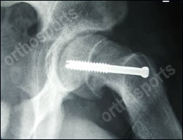
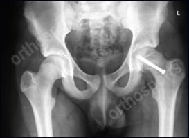
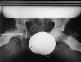
Treatment of slipped epiphysis involves putting a screw up the neck of the femur, across the growth plate and into the head to prevent further slipping and encourage the growth plate to close.

Lateral Hip X-ray

AP Pelvis X-ray

Frog Leg Lateral X-ray
All images above show a screw preventing further slippage.
If there is a severe deformity after the growth plate has been stabilized it may be necessary to change the shape of the upper femur to improve the mechanics of the hip joint and decrease or delay the onset of premature osteoarthritis. This is an osteotomy.
Osteoarthritis in adult life is a complication of slipped epiphysis because of the altered mechanics of the hip joint secondary to deformity of the bone after the slip.
Any adolescent with knee or thigh pain and a limp should have a frog leg lateral hip x-ray to look for a slipped epiphysis.
