Orthosports GP Newsletters

Welcome to our newsletters. In this series you will find short but illustrative clinical cases and examination techniques to keep you up to date with common Orthopaedic presentations you are likely to see in your office.
Shoulder Examination
Shoulder Examination Part 1:
Presented by: Dr Doron Sher
To view the Shoulder Examination Video please Click Here
Shoulder pain is common and can be difficult to diagnose accurately. It is helpful to remember that certain diagnoses are more common in certain age groups.
Across the board, impingement is by far the most common diagnosis but in the younger age groups (20-40) one should consider instability both as it’s own diagnosis and as a cause for secondary impingement. In the 30-50 year age group remember adhesive Capsulitis (which unfortunately is overdiagnosed) as well as impingment; and in the 50+ age group add arthritis to the list above.
As with all joints a thorough history is required to appropriately direct your clinical examination. Ask about the patient’s age, hand dominance, sport and work activities. Does the injury prevent or interfere with work, hobbies and sport?
The location and nature of the pain, instability, stiffness, locking, catching and swelling are all critical questions.
Pain from the shoulder is often felt in the upper arm and night pain is common.
Stiffness or loss of motion is seen with adhesive capsulitis, arthritis and posterior glenohumeral dislocation. Pain or inability to throw suggests anterior glenohumeral instability. Pain at the top of the shoulder may be AC joint arthritis. A history of a fall, or pain with lifting, may indicate a rotator cuff tear and may be associated with bruising in the upper arm (which could also indicate a fracture).
Clinical examination requires inspection, palpation, evaluation of range of motion and provocative testing. Always remember to check the neck and elbow and look for neurological problems. Bones, muscles and soft tissue structures can all be injured around the shoulder.
The rotator cuff is made up of 4 muscles:
The supraspinatus, infraspinatus, teres minor and subscapularis, with the supraspinatus being the most commonly injured muscle.
INSPECTION: The shoulders and arms should be properly exposed to look for swelling, asymmetry, muscle atrophy, scars and bruising. Loss of shoulder roundness may be from a dislocation or chronic muscle wasting. Scapular winging and poor scapulothoracic rhythm can indicate a nerve injury or be the cause of secondary impingement. Wasting of the supraspinatus or infraspinatus makes you suspicious of a rotator cuff tear, suprascapular nerve entrapment or neuropathy.
PALPATION: Palpation should include examination of the acromioclavicular and sternoclavicular joints, the cervical spine and the biceps tendon. The glenohumeral joint, coracoid process, acromion and scapula should also be palpated for any tenderness and deformity.
RANGE-OF-MOTION TESTING: Always compare the two shoulders with both active and passive range of motion. Forward elevation, external and internal rotation and abduction are the most useful movements. A difference in active and passive motion is usually caused by a rotator cuff tear. Loss of some motion can be caused by impingement, calcific tendonitis and chronic instability. A global loss of motion is caused by arthritis, chronic dislocations, fractures and massive rotator cuff tears.
STRENGTH TESTING: When testing the rotator cuff be sure to compare the painful side to the unaffected side to detect subtle differences in strength and motion. True weakness should be distinguished from weakness that is due to pain. A patient with subacromial bursitis with a tear of the rotator cuff often has objective rotator cuff weakness caused by pain when the arm is positioned in the arc of impingement, however the rest of the rotator cuff examination may be normal.
The supraspinatus can be tested by having the patient abduct the shoulders to 90 degrees in forward flexion with the thumbs pointing downward. The patient then attempts to elevate the arms against examiner resistance. This is often referred to as the “empty can” test.
Next, with the patient’s arms at the sides, the patient flexes both elbows to 90 degrees while the examiner provides resistance against external rotation. Weakness of external rotation almost always indicates a rotator cuff tear.
Subscapularis function is assessed with the lift-off test. The patient rests the dorsum of the hand on the back in the lumbar area. Inability to move the hand off the back by further internal rotation of the arm suggests injury to the subscapularis muscle. A modified version of the lift-off test is useful in a patient who cannot place the hand behind the back. In this version, the patient places the hand of the affected arm on the abdomen and resists the examiner’s attempts to externally rotate the arm.
Injections can be used for diagnosis and treatment. If a subacromial injection relieves pain and restores motion and strength the patient usually has impingment rather than a rotator cuff tear.
AC joint injections are useful to deal with superior shoulder pain (please refer to the “Teaching” Section of our website and go to “Injection Techniques”).
Shoulder Examination Part 2:
Presented by Dr Todd Gothelf
Examination of the Shoulder for Rotator Cuff Disease:
ANATOMY OF THE ROTATOR CUFF: Rotator cuff tears most commonly begin with the supraspinatus, resulting in weakness in external rotation. Subscapularis tears result in internal rotation weakness.

The physical examination for a rotator cuff tear will serve to isolate out these muscles to test their function. A patient with an intact rotator cuff will be able to resist the examiner’s force during strength testing. When a patient is unable to resist, weakness is noted.
If the patient is in pain a local anaesthetic INJECTION into the subacromial space can numb the area, eliminating pain so that rotator cuff power can be accurately tested.
Xrays are necessary to rule out arthritis and assess the acromion and joint position. A MRI arthrogram is the gold standard to assess a rotator cuff tear. Ultrasound is not a useful tool for surgical decision making.
WHERE IS THE SOURCE OF PAIN? Shoulder pain may originate from pathology in the neck. Pain is REFERRED to the shoulder and can make diagnosing the source difficult.
Examination of the shoulder begins with the neck. The patient is asked to extend the neck and tilt from side to side. This is known as the SPURLING MANOUEVRE. This movement puts pressure on the nerves exiting the neck and will reproduce the pain if the problem is in the neck rather than the shoulder.
Rotator cuff tear pain is elicited with movements of the glenohumeral joint.
Impingement tests known as the NEER and HAWKINS sign are used to demonstrate this pain. These help to diagnose impingement, calcific tendinitis, adhesive capsulitis, or rotator cuff tears, but they do not prove the presence of a rotator cuff tear.
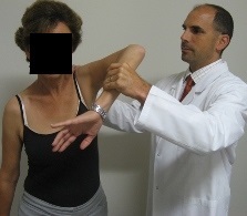
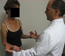
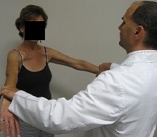
NEER SIGN – The arm is slightly internally rotated with the thumb pointed down. The shoulder is forward flexed with the elbow straight. Pain occurs at the acromion with a positive sign.
HAWKINS SIGN – With the shoulder forward, the elbow is flexed to 90 degrees and the arm is further internally rotated. A positive test produces pain near the acromion. (Image above)
HOW ARE DIFFERENT SHOULDER DIAGNOSES DIFFERENTIATED?
RANGE OF MOTION TESTING – Testing Active and Passive motion helps to differentiate a rotator cuff tear from adhesive capsulitis or glenohumeral arthritis. PASSIVE motion of the shoulder is generally maintained with a rotator cuff tear and restricted in the other entities. ACTIVE motion will be limited as the torn rotator cuff limits the patient’s ability to move the shoulder in all ranges of motion. If the problem has been present for some time secondary stiffness can occur.
STRENGTH TESTING – The rotator cuff provides power to shoulder movement, and a tear will result in weakness. The earliest signs of weakness will be evident in an external rotation strength test (Image above). With the arms placed at the sides and forearms forward, the patient is asked to externally rotate against the resistance of the examiner. The examiner pushed inward with both arms, and a difference in power is noted. The arms are placed about 30 degrees from straight to the sides, elbows straight and thumbs pointing downward (Empty Can Test) Any weakness is noted (Image above).
DROP ARM TEST – With a very large tear, the patient may lose the ability to lift the arm against gravity. The arms are held in the plane of the scapula at 90 degrees, and the patient is asked to slowly lower their arms to the side. With a large rotator cuff tear the patient will have trouble moving slowly, and the arm will quickly drop.
HORNBLOWER’S SIGN – This specific test demonstrates a very large rotator cuff tear. The arm is abduction 90 degrees and the elbow flexed. The patient is unable to keep the arm externally rotated, and it falls forward in a position that appears like a hornblower’s position. This test isolates infraspinatus, which is the main driver of external rotation. A positive test confirms a massive tear and significant loss of function of the rotator cuff.
SUBSCAPULARIS TEARS – These tears result in loss of INTERNAL rotation strength, and should be looked for as they can occur in isolation.
LIFT OFF TEST – The hand is placed on the lower back, palm outward, and the patient is asked to lift the hand off the back. Inability to do so represents weakness of the subscapularis.
If a direct anterior drawer is obtained, the ACL will be torn. However, for this sign to be elicited, peripheral structures such as the medial meniscus or the menisco-tibial ligament must also be damaged. This ligament forms a wedge, in 90° flexion, preventing anterior tibial translation. The finding of an anterior drawer is conclusive evidence of an ACL tear. However, not every ACL tear will be associated with a positive anterior drawer test.
Shoulder Examination Part 3:
Presented by Dr Jerome Goldberg
the patient with Shoulder instability:

Specifically if the arm is in abduction and external rotation when the patient has the sensation of instability, the instability then is in an anterior direction. If the arm is adducted and internally rotated the instability is posterior. Apart from a frank dislocation or subluxation the patient may have a subtle instability and complain of “the arm going dead” with forced abduction and external rotation of the arm such as when going into a tackle, throwing or serving at tennis. Beware of the patient who complains of instability with the arm by the side. Patients who are hypermobile or have generalised ligamentous laxity are more prone to have instability than others.
Remember the examination concept of look, feel, and move.
Look for wasting of the shoulder girdle muscles, rupture of the long head of biceps and any scarring. Feel the whole shoulder girdle and determine the tender points. In particular, tenderness of the joint lines, anterior and/or posterior, often occur in instability.
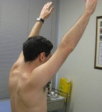
Examine the patient elevating the arms in forward flexion from both the back and the front. Observe any abnormality of rhythm and compare the injured to the non injured side.
Specifically observe the movement from behind and watch the scapula move comparing one side to the other. Although uncommon if the scapula wings as the patient brings the arm down one should suspect a scapula dysrhythmia, as a potential cause of the problem. Check the patient has a full range of motion.
Look for ligamentous laxity by checking if the thumb can reach the back of the forearm.
The apprehension signs are designed to determine whether the shoulder is unstable. The aim is to put the shoulder in the provocative position for the dislocation and reproduce the patient’s symptoms without actually dislocating the shoulder. For an anterior dislocation the shoulder should be put in abduction and external rotation, and for a posterior instability the direction of force should be adduction and internal rotation. This test can generally be done with the patient sitting on an examination couch but in the larger patient it is often easier to have the patient in the supine position.
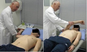
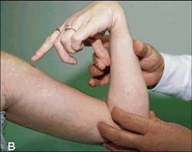
Inferior instability can be determined by pulling the arm inferiorly and reproducing a sulcus sign.
If you suspect an instability then plain xrays are required to exclude fractures of the glenoid in particular (the so called bony Bankart lesion) or glenoid dysplasia. Ultrasounds are of little value. If surgery is contemplated the patient requires an MRI with intraarticular contrast. An MRI without contrast is not as helpful.

Shoulder Injury related to vaccine administration
Welcome to Orthosports News. This month Dr Doron Sher discusses Shoulder Injury related to Vaccine administration.Please feel free to send your questions to education@orthosports.com.au A JAB
GP News – August 2021
This month Dr Doron Sher presents a Knee Examination series that you may find useful for patients presenting in your practice requiring a knee assessment.
Painful Ankle
Case Study: Painful Ankle Presented by: Dr John Negrine A 39 Year old man presents with a painful ankle: He strenuously denies any history of
Previous Newsletters:

Welcome to Orthosports GP Online Newsletter. Below you will find two articles which we trust you will find useful in your practice.
Article 1:
Article 2:


